-
1 of 253523 objects
The fetus in the womb; sketches and notes on reproduction c.1511
Recto: Red chalk and traces of black chalk, pen and ink, wash. Verso: Pen and ink, with some offset red chalk | 30.4 x 22.0 cm (sheet of paper) | RCIN 919102

Leonardo da Vinci (1452-1519)
The fetus in the womb; sketches and notes on reproduction c.1511

Leonardo da Vinci (1452-1519)
The fetus in the womb; sketches and notes on reproduction c.1511


-
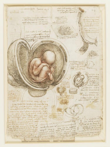
A drawing of a fetus in utero, with the uterus opened out; a smaller sketch of the same, and of details of the placenta and uterus; a diagram demonstrating binocular vision; notes on embryology, on relief in painting and on mechanics.
Throughout his anatomical career Leonardo strove for objectivity, and colour is usually absent in the drawings themselves (if not the paper). But in his late embryological drawings he was clearly moved by the ineffable mystery of life, as he had been at the start of his anatomical researches twenty-five years before, and this sheet, perhaps the best known of all Leonardo’s anatomical drawings, makes striking use of red chalk and dense curved hatching to evoke the coiled potential of the child in the womb. Leonardo’s renewed concern with the immaterial aspects of the body is expressed in the note to lower left:
"In this child the heart does not beat and it does not breathe because it rests continually in water, and if it breathed it would drown. And breathing is not necessary because it is vivified and nourished by the life and food of the mother... And one and the same soul governs these two bodies, and desires, fears and pains are common to this creature as to all other animated parts. From this it arises that a thing desired by the mother is often found imprinted on those parts of the infant that have the same qualities in the mother at the time of her desire; and a sudden terror kills both mother and child. Therefore one concludes that the same soul governs and nourishes both bodies."
The principal drawing shows the fetus in ‘complete breech’ position with the legs crossed, though here the left heel is not pressed against the perineum as in RCIN 919101. The umbilical cord is wrapped around the fetus’s legs, but its connection to the placenta is not seen; in the left margin is an ovary and associated vessels. In the geometrical diagram at centre right Leonardo implies that the weight of the fetus’s head might tip it over in the uterus for normal head-first delivery, by analogy with an eccentrically weighted sphere rolling a little way up an incline. The small sketches below centre envisage the uterine membranes unfurling one by one like the petals of a flower, and in the main drawing this allows Leonardo to show the membranes in cross-section. Particular attention is given to the multiple cotyledenous placenta (derived from Leonardo’s dissection of a cow recorded on 919055), whose interdigitations are shown in the details at upper right. Leonardo never discovered that the human has a single, discoidal placenta, and his ongoing reliance on bovine material is indicated by the note ‘get the secondine [fetal membranes] of calves when they are born and note the shape of the cotyledons’.
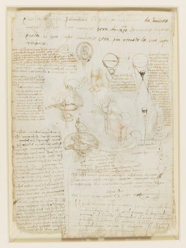
On the verso: sketches of a fetus in utero, and of a fetal liver, stomach and umbilical vein; sketches of a chick embryo and membranes; notes on reproduction, and on light and shade. In addition to the notes in Leonardo's hand, there are further notes in two hands - the top four lines by his pupil Francesco Melzi, the bottom right portion by an unknown hand, perhaps Leonardo's anatomical associate Marcantonio della Torre (d.1511).
Text adapted from M. Clayton and R. Philo, Leonardo da Vinci: Anatomist, London 2012Provenance
Bequeathed to Francesco Melzi; from whose heirs purchased by Pompeo Leoni, c.1582-90; Thomas Howard, 14th Earl of Arundel, by 1630; probably acquired by Charles II; Royal Collection by 1690
-
Creator(s)
Acquirer(s)
-
Medium and techniques
Recto: Red chalk and traces of black chalk, pen and ink, wash. Verso: Pen and ink, with some offset red chalk
Measurements
30.4 x 22.0 cm (sheet of paper)
Other number(s)