-
1 of 253523 objects
The fetus, and the muscles attached to the pelvis (recto); studies of the fetus, related internal organs, and the arm (verso) c.1511
Recto: Red and black chalks, pen and ink, wash. Verso: Pen and ink | 30.4 x 21.3 cm (sheet of paper) | RCIN 919101

Leonardo da Vinci (1452-1519)
The fetus, and the muscles attached to the pelvis (recto); studies of the fetus, related internal organs, and the arm (verso) c.1511

Leonardo da Vinci (1452-1519)
The fetus, and the muscles attached to the pelvis (recto); studies of the fetus, related internal organs, and the arm (verso) c.1511


-
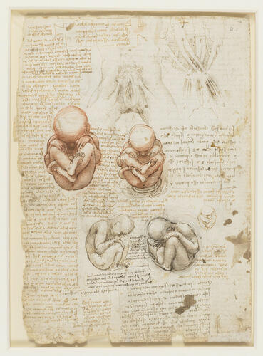
Recto: studies of the female genitalia and abdominal muscles; four drawings of fetuses, and a small sketch of a fetus in the uterine position; notes on the drawings.
Leonardo was fascinated by how a fetus could fit into the uterus of a woman – here he states that ‘the length of a child when it is born is usually one braccio [c. 60 cm]’ but ‘experience in the dead shows [the uterus] to be a quarter of a braccio in its greatest length in a woman’. He repeatedly drew the fetus curled up to occupy the smallest space possible, and in the long note in the left margin he tries to compare the size of the human uterus with that of the cow and the horse, in proportion to their bodies.
By curling the fetus up in this manner, Leonardo noted that the heel is pressed against the perineum. He therefore suggests that:
"During a great part of the period of life of the fetus its urination is through the umbilicus. And this happens because the heel of the right foot is comes between the anus and the penis and shuts off the passage of all urine. Nature has provided for this by making a channel in the fundus of the bladder [the urachus, the fibrous remnant of the allantois] through which urine goes from the bladder to the umbilicus and from the umbilical cord to the mouth of the uterus."
The two pen drawings at the top of the page examine the supposed action of muscles attached to the pubis. The external and internal abdominal oblique muscles are broad flat sheets laid over one another, attached to the ribs above and inserting partly on the iliac crest and partly in aponeuroses which meet at the linea alba, the mid-line of the abdomen. Their muscle fibres run diagonally, those of the external obliques roughly perpendicular to those of the internal obliques, and it would appear to be this arrangement that Leonardo has tried to record (he has not shown the rectus abdominis muscles). He has however rendered the muscles as a series of long thin structures, attached to the pubis below and to four unidentifiable points above, and crossing over at the mid-line, which they do not do (the same arrangement was sketched more than twenty years earlier on 912609). Below the pubis Leonardo has shown the adductor muscles of the thigh, and his accompanying note states that the opposing pulls of the abdominal and adductor muscles stabilise the pubis; the reality is the reverse, with the pubis serving as an anchor point for the actions of the muscles.
Text adapted from M. Clayton and R. Philo, Leonardo da Vinci: Anatomist, London 2012.
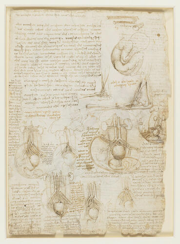
Verso: sections of the umbilical cord; a fetus in profile to the right; drawings of the tendons and bones of the right elbow and forearm; seven drawings of fetal viscera, blood vessels and the umbilical cord; notes on the drawings.Provenance
Bequeathed to Francesco Melzi; from whose heirs purchased by Pompeo Leoni, c.1582-90; Thomas Howard, 14th Earl of Arundel, by 1630; probably acquired by Charles II; Royal Collection by 1690
-
Creator(s)
Acquirer(s)
-
Medium and techniques
Recto: Red and black chalks, pen and ink, wash. Verso: Pen and ink
Measurements
30.4 x 21.3 cm (sheet of paper)
Other number(s)