-
1 of 253523 objects
Recto: The vulva and anus. Verso: The male and female reproductive systems c. 1508
Pen and ink over black chalk | 19.1 x 13.8 cm (sheet of paper) | RCIN 919095
-
A folio from Leonardo's 'Anatomical Manuscript B'.
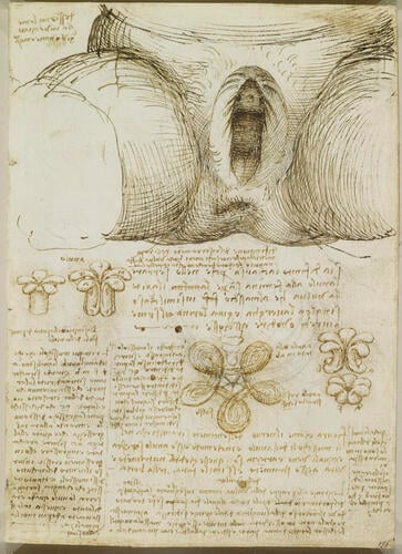
Recto: A drawing of the external genitalia and vagina, with notes; notes on the anal sphincter, with diagrams of the suggested arrangement of its fibres and its mode of action.
The principal drawing shows the vulva of a multiparous woman, with the labia majora separated to show the urethral orifice and the transverse rugae of the vagina; the labia minora are not evident. The urethral orifice is almost papilliform, and as this individual had given birth multiple times there may have been some prolapse, a weakness in the musculo-tendinous floor of the pelvis which holds the bladder.Leonardo was interested in how the ‘six gaps in the skin’ could open and close – ‘the eyes, nostrils, mouth, vulva [or] penis and anus, and the heart though it is not in the skin’. He maintained, quite correctly, the principle that a muscle can only pull and not push, but it did not occur to him that a muscle could be ring-shaped rather than linear, and so he failed to comprehend the nature of sphincter muscles. Here he posited that the anus consisted of a ring of five separate muscles, each of which in contraction would increase in thickness, so closing the anus. It was unclear even to Leonardo himself why there should be five such muscles: he asked ‘why the muscles of the anus are odd in number; and if this disparity were necessary, why were three or seven not chosen rather than five?’. Possibly he derived this figure from the number of ‘pyramidal wrinkles’ visible at the anus in his subject – on 919055v Leonardo states that ‘the skin which forms the covering of muscles which pull always directs its wrinkles to the place where the cause of movement is’.
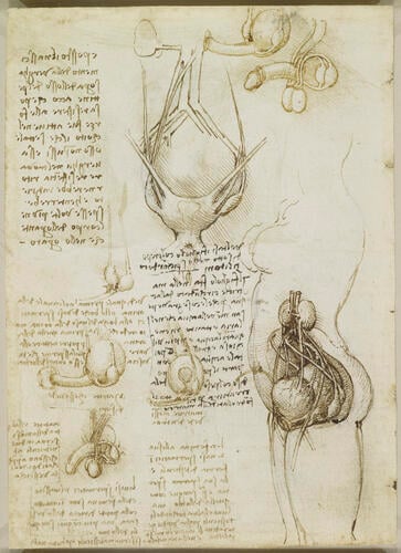
Verso: The left side of a standing female showing the uterus in early pregnancy; an anterior view of the female genito-urinary system; six drawings of the male and female genitalia.
Here Leonardo attempts to analyse the similarities between the male and female generative organs:
"The female has her two spermatic vessels in the form of testicles, and her sperm is first blood, like that of the male. But in both one and the other [female and male], on reaching the testicles it takes on generative power... Neither in the one nor the other is it kept in the testicles, but in one in the uterus and in the other, that is the male, in the two ventricles [seminal vesicles] attached to the back of the bladder."
It is thus clear that Leonardo regarded both male and female as contributing equally to conception (cf. 919097); he calls the ovaries ‘testicles’, and states that they also form sperm from blood and send it to the uterus. But he does not try to impose these homologies too strictly. Although in these drawings the seminal vesicles are of the same shape and in the same position as the ovaries, he is clear that it is the testicles that are the source of sperm, which is simply held in the vesicles. He has also now discovered that the urethra is the only channel in the penis, and that the ejaculatory ducts issue into the urethra.
The external view of the uterus at upper centre, however, shows that Leonardo still had a poor understanding of the region. Three structures are shown attached at either side; these may be (from top to bottom) a uterine ligament, seen in a variety of configurations in Leonardo’s drawings of the period (cf. 912281); the mythical vessel thought to carry menstrual blood during pregnancy to the breast for conversion into milk (cf. 919097); and the ovary, with the ovarian vein at its upper end and discharging ‘female sperm’ (alluded to in the passage quoted above) from its lower end into the uterus. The drawing at lower right tries to make three-dimensional sense of these supposed structures. Most clearly visible is the bladder at the front of the body, with the ureters passing from the kidneys and what is presumably the superior vesical artery emanating from the internal iliac artery . Behind the bladder is the uterus, with the left ovary joined to a vessel that can be traced upwards to the region of the kidneys (the left ovarian vein does indeed connect to the left renal vein); and the ‘menstrual blood vessels’ loop upwards from both sides of the uterus to peter out in front of the kidneys. A further blood vessel seems to pass down towards the front of the bladder, from where another passes back up to the umbilicus; and the colon can be seen curving around behind the uterus, terminating in a dark patch of ink at the anus.
Text from M. Clayton and R. Philo, Leonardo da Vinci: Anatomist, London 2012Provenance
Bequeathed to Francesco Melzi; from whose heirs purchased by Pompeo Leoni, c.1582-90; Thomas Howard, 14th Earl of Arundel, by 1630; Probably acquired by Charles II; Royal Collection by 1690
-
Creator(s)
Acquirer(s)
-
Medium and techniques
Pen and ink over black chalk
Measurements
19.1 x 13.8 cm (sheet of paper)
Other number(s)

