-
1 of 253523 objects
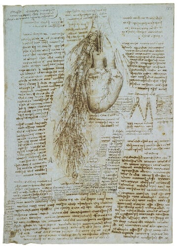
The heart, bronchi and bronchial vessels (recto); A sketch of the heart and great vessels (verso) c.1511-13
Recto: Pen and ink on blue paper. Verso: Black chalk on blue paper | 28.8 x 20.3 cm (sheet of paper) | RCIN 919071

Leonardo da Vinci (1452-1519)
The heart, bronchi and bronchial vessels (recto); A sketch of the heart and great vessels (verso) c.1511-13

Leonardo da Vinci (1452-1519)
The heart, bronchi and bronchial vessels (recto); A sketch of the heart and great vessels (verso) c.1511-13


-
Recto: a drawing of a bovine heart, the great vessels and the bronchial tree; details of the trachea and the effects of respiration on it; numerous notes on the action of the heart.
The drawing is a view from behind of the heart, bronchi and bronchial vessels of an ox, the subject of most of Leonardo’s heart dissections. The coronary sinus and circumflex branch of the left coronary artery are seen in the posterior left atrioventricular groove, with the middle cardiac vein and posterior interventricular artery descending in the posterior interventricular groove. Leonardo shows the branching of the bronchi, with their walls formed of regular cartilaginous tubes: in reality the trachea has evenly spaced ‘C’ shaped cartilages within its wall (as Leonardo had described on 919050v), the primary bronchi have large plates, and the amount of cartilage decreases through the secondary and tertiary bronchi. The bronchi are accompanied throughout by ramifying veins and arteries which Leonardo has attempted to represent as paired; he shows at least two left and one right bronchial arteries arising from the arch of the aorta, but the drainage of the bronchial veins is not indicated. At centre right are details of a ‘trachea of medium size’ (to the right) and a ‘smallest trachea uninflated and inflated, which is doubled in capacity in its expansion’ – Leonardo used the term ‘trachea’ to refer to all the passages of the respiratory tract.
The extensive notes include the following passage:
Whether air penetrates into the heart or not
To me it seems impossible that any air can penetrate into the heart through the trachea [ie. bronchi], because if one inflates [the lung], no part of the air escapes from any part of it. And this occurs because of the dense membrane with which the entire ramification of the trachea is clothed. This ramification of the trachea as it goes on divides into the most minute branches together with the most minute ramification of the veins which accompany them in continuous contact right to the ends. It is not here that the enclosed air is breathed out through the fine branches of the trachea and penetrates through the pores of the smallest branches of these veins. But concerning this I shall not wholly affirm my first statement until I have seen the dissection which I have in hand.
Leonardo thus refutes the traditional belief that air passes from the lungs into the heart (as illustrated in 919104v), and indeed that air mixes directly with blood anywhere in the cardiopulmonary system. That this refutation is both experimental and provisional demonstrates Leonardo’s maturity as a scientist. Had he omitted the word ‘not’ four lines from the end of the above passage, Leonardo would have provided a perfect description of gaseous exchange in the alveoli; but he had, of course, no knowledge of this process, and he maintained to the end of his life a belief that the lungs existed to cool the blood heated by its turbulence in the chambers of the heart.
(Text from M. Clayton and R. Philo, Leonardo da Vinci: Anatomist, London 2012)
*
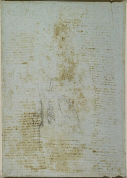
Verso: a sketch of a human heart and the main vessels.
Provenance
Bequeathed to Francesco Melzi; from whose heirs purchased by Pompeo Leoni, c.1582-90; Thomas Howard, 14th Earl of Arundel, by 1630; probably acquired by Charles II; Royal Collection by 1690
-
Creator(s)
Acquirer(s)
-
Medium and techniques
Recto: Pen and ink on blue paper. Verso: Black chalk on blue paper
Measurements
28.8 x 20.3 cm (sheet of paper)
Other number(s)