-
1 of 253523 objects
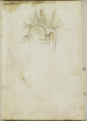
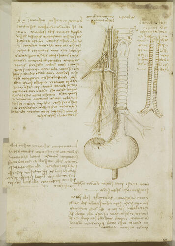
Recto: The arteries of the shoulder. Verso: The distribution of the right vagus and right phrenic nerves c. 1508
Pen and ink over black chalk | 19.3 x 13.3 cm (sheet of paper) | RCIN 919050
-
A folio from Leonardo's 'Anatomical Manuscript B'.
Recto: a drawing of the large arteries of the neck, thorax and right upper arm, with a note that they are those of an old man.
Verso: a study of the larynx, trachea, oesophagus and stomach, with the phrenic and right vagus nerves, the brachial plexus and some blood vessels; to the right, the trachea seen from behind; notes on the drawings and on the brain. The principal drawing (labelled 'del vechio', 'of the old man'; see RCIN 919027) shows the trachea down to its bifurcation into the primary bronchi, below which the oesophagus is visible, leading to the stomach. Leonardo accurately follows the course of the right vagus nerve down the right side of the trachea, then along the oesophagus and finally around the lesser curvature of the stomach. Immediately to the left of the upper part of the nerve are the internal jugular vein and carotid artery, running vertically down to join the subclavian vein and artery and form the right brachiocephalic vein and artery, respectively. Further to the left is the gently curving external jugular vein, with no accompanying artery.
Behind these vessels can be seen the brachial plexus, with the right phrenic nerve (the motor supply of the diaphragm) running from n to m, and accompanied by the pericardiacophrenic vessels, exaggerated in size. At about the mid-point of the trachea, the right recurrent laryngeal nerve can be seen branching off from the right vagus nerve and looping around the brachiocephalic vessels to travel upwards and innervate the trachea and larynx. Leonardo has confused the topography of this area: in reality the brachiocephalic artery and its branches lie behind their venous corollaries; the vagus nerve passes in front of the arterial branches, and the recurrent laryngeal nerve loops backwards underneath them.
The note below reminds Leonardo to ‘observe in what way the reversive [recurrent laryngeal] nerves give sensation to the rings of the trachea, and which muscles give movement to these rings in order to produce a deep, medium or high-pitched voice’. The diagram to the right shows a longitudinal section of the trachea and proximal bronchi, with the cartilaginous structures commonly referred to as tracheal ‘rings’. These are in fact ‘C’ shaped and incomplete towards the spine; Leonardo suggests that this is ‘to give room for the food [in the oesophagus] between themselves and the bone of the neck.’
Text from M. Clayton and R. Philo, Leonardo da Vinci: Anatomist, London 2012
Provenance
Bequeathed to Francesco Melzi; from whose heirs purchased by Pompeo Leoni, c.1582-90; Thomas Howard, 14th Earl of Arundel, by 1630; Probably acquired by Charles II; Royal Collection by 1690
-
Creator(s)
Acquirer(s)
-
Medium and techniques
Pen and ink over black chalk
Measurements
19.3 x 13.3 cm (sheet of paper)
Other number(s)

