-
1 of 253523 objects
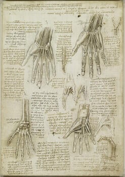
The bones, muscles and tendons of the hand c.1510-11
Black chalk, pen and ink, wash | 28.8 x 20.2 cm (sheet of paper) | RCIN 919009
-
A folio from Leonardo's 'Anatomical Manuscript A'.
Recto: five studies of right hands, showing bones and muscles; six smaller drawings of digits; notes on the drawings.
These magnificent drawings are among the high points of Leonardo’s career as an anatomist. They demonstrate with complete clarity the mechanical structure of the hand, not stripping it down as in a dissection but building it up in the manner of an engineer, and following in part the list of depictions given on the verso of the sheet. Leonardo begins at lower left with the bones (labelled 1st), then adds the deep muscles and tendons of the palm and wrist at lower right (labelled 2nd), the first layer of tendons at upper left (3rd), and the second layer of tendons at upper right (4th). Two further drawings, adding the nerves and then the blood vessels, are labelled 5th and 6th on RCIN 919012v.The bones of the wrist are a little confused in the first drawing, and are more clearly rendered on the verso of the sheet. A thread between the radius and the first metacarpal bone of the thumb is possibly the radial or lateral collateral ligament, or a tendon of abductor pollicis longus or extensor brevis. In the second drawing the bones are clothed in the deepest muscle and tendon. We see the thenar and hypothenar muscle compartments, controlling the thumb and little finger respectively, and the deep transverse metacarpal ligament spanning the knuckles to keep them from separating. The carpal tunnel is open, and pronator quadratus is shown connecting the radius to the ulna.
At upper left Leonardo adds the tendons of flexor digitorum profundus, running from the muscles on the anterior side of the ulna, through the carpal tunnel beneath the transverse carpal ligament (here represented by two threads), to the fingertips. The tendons of flexor digitorum superficialis are added at upper right, along with the ulnar and median nerves, reflected to the sides of the wrist, and some of the fibrous sheaths and annular ligaments that hold the tendons in place and thus allow the bending of the fingers, as demonstrated at lower right. As their names indicate, flexor digitorum profundus lies below superficialis, but its tendons attach further down the fingers, and consequently the tendons of profundus penetrate those of superficialis, as shown in the diagram at upper centre (this gap in the tendon of superficialis is named ‘Camper’s chiasm’ after the anatomist Peter Camper, who described it in 1760-62; it should perhaps be renamed ‘Leonardo’s chiasm’). Leonardo was entranced by the elegance of this arrangement, and surrounded that detail with the note ‘Provide that the book on the elements of mechanics, with its practice, comes before the demonstration of the movement and force of man and other animals; and by these means you will be able to prove all your propositions.’
*
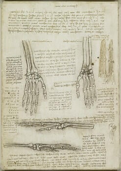
Verso: studies of the bones of a right hand, and of individual fingers; studies of the skeleton of a right hand; a clenched fist; notes on the drawings. At the top of the page Leonardo sets out the sequence of depictions of the hand that he intended to provide:
"The first demonstration of the hand will be of its bones alone. The second, of the ligaments and various interconnections of tendons that join them together. The third will be of the muscles that arise on these bones. The fourth will be of the first tendons that are placed on these muscles, and give movement to the fingertips. The fifth will show the second rank of tendons, which move all the fingers and terminate on the penultimate piece of bone of the fingers. The sixth will show the nerves that give sensation to the fingers of the hand. The seventh will show the veins and arteries that give nourishment and spirit to the fingers. The eighth and last will be the hand clothed with skin, and this will be drawn for an old man, a young man and a child; and for each will be given the measurements of length, thickness and width for each of its parts."
These intentions were mostly carried out, here and on the recto of the sheet and 919012v, though Leonardo did not, so far as we know, make a sequence of drawings of subjects of different ages. We also have no clear depiction of the second stage, showing the ligaments connecting the bones, though the drawing at lower left of the recto (which seems at first glance essentially to duplicate that at upper right here) may initially have been intended to be such a drawing.
The two largest drawings here give palmar (right) and dorsal (left) views of a right hand; below are views from either side, lateral then medial, with the thumb slightly lowered. Leonardo thus gives four orthogonal views of the same stage of dissection. But his ambitions were endless, and having made these four drawings he then stated his wish to depict each individual bone in four aspects. The magnitude of this task is emphasised by the note at centre left, which enumerates the 27 bones of the hand (identified by letters and numbers) – thus 108 individual drawings of those bones alone.
The drawings in the right margin show all the different structures coursing into the finger. Leonardo identifies the tendon of extensor digitorum, which straightens the finger; ‘the vein that nourishes the finger’; the nerve; ‘the vein that gives vital spirit to the finger’ (i.e. the artery); and the tendon of flexor digitorum profundus, which bends the finger.
Text from M. Clayton and R. Philo, Leonardo da Vinci: Anatomist, London 2012Provenance
Bequeathed to Francesco Melzi; from whose heirs purchased by Pompeo Leoni, c.1582-90; Thomas Howard, 14th Earl of Arundel, by 1630; probably acquired by Charles II; Royal Collection by 1690
-
Creator(s)
Acquirer(s)
-
Medium and techniques
Black chalk, pen and ink, wash
Measurements
28.8 x 20.2 cm (sheet of paper)
Other number(s)

