-
1 of 253523 objects
The muscles of the arm, and the veins of the arm and trunk (recto); The muscles of the shoulder, arm and neck (verso) c.1510-11
Black chalk, pen and ink, wash | 28.9 x 19.9 cm (sheet of paper) | RCIN 919005

Leonardo da Vinci (1452-1519)
The muscles of the arm, and the veins of the arm and trunk (recto); The muscles of the shoulder, arm and neck (verso) c.1510-11


-
A folio from Leonardo's 'Anatomical Manuscript A'.
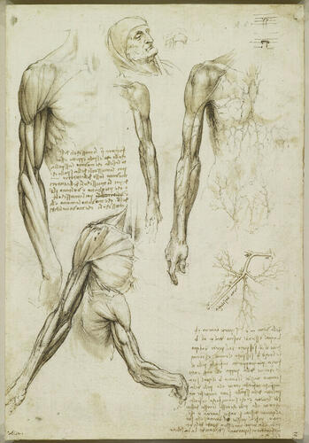
Recto: studies of musculature in the right arm; the head of an old man; a diagrammatic drawing of the jaw; a study of a left arm, showing the veins; veins of the body and the right arm; a detailed drawing of the basilic vein and tributaries; notes on the drawings.
The elderly and apparently dead man depicted at the top of the sheet was presumably the subject of some of the dissection studies here and elsewhere in Manuscript A. To the right of the head is a sketch of his jaw and neck, with the tongue and hyoid sunken in the flaccidity of recent death. The three consummately elegant studies of the upper limb down the left side of the page, however, give the old man the musculature of Apollo. With a few dabs of wash Leonardo has captured the shimmer of the deep fascia, the connective tissue surrounding and interpenetrating the muscles. As was usual throughout the Renaissance, the corpse is posed as if in action, the muscles imbued with the tension of life. Leonardo has not resorted to diagrammatic devices (such as fenestrating pectoralis major, as is seen repeatedly throughout the manuscript), and all the muscles are identifiable.
The large drawing at upper right shows the trunk with only the skin removed. The cutaneous veins of the arm – cephalic, basilic and median cubital – are visible, and on the trunk can be seen the lateral thoracic vein, superficial epigastric veins, tributaries of the internal thoracic vein and possibly a thoracoepigastric vein. Below is a more detailed study of the basilic vein as it enters the axillary vein and then in turn the subclavian vein. The identities of the other veins in this detail are not certain as there is a large amount of venous drainage in the axillary region, but the cephalic, subscapular, lateral thoracic, thoracoepigastric and thoracodorsal veins are probably all present.
*
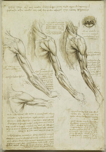
Verso: four studies of the right shoulder and arm; a slight study of the elbow; a front view of an open mouth, showing molar teeth, palate, uvula and throat; notes on the drawings.
This page together with 919008v form a sequence of eight drawings in which Leonardo turned the shoulder and arm from a fully anterior view (at far right) to a fully posterior view. The small star-shaped diagram on 919008v and accompanying note at lower right explain Leonardo’s intention of depicting the arm through 360° from eight aspects, but the two pages together provide an even more finely divided set of depictions, eight aspects in 180°. There are, as always, a few idiosyncrasies, but the drawings and notes reveal a profound understanding of the muscles of the shoulder and upper arm. Medical illustration has never produced images to surpass those on this sheet, and had Leonardo published his researches, as was clearly in the forefront of his mind at this time, the results would have been truly groundbreaking.
Most of the superficial muscles of the arm and shoulder can be identified. While pectoralis major is shown as a single muscle, the deltoid (forming the rounded upper contour of the shoulder) is as usual depicted as a compound structure comprising distinct sets of fibres. Likewise, the superior portions of the trapezius muscle, running upwards from the spine of the scapula, appear to be separate.
Towards the wrist, at centre right of the present page, Leonardo has labelled the tendons of the abductor pollicis longus and extensor pollicis brevis muscles, both of which act on the thumb, reminding himself to remove the covering muscle (the supinator) to find their origins. He goes on to note:
"Do the same for all the muscles, leaving each one on its own, naked on the bone, so that besides seeing its beginning and end, it is shown in what way it moves the bone to which it is dedicated; and of this, the scientific explanation [ragione scientificha] is to be provided with lines alone [ie. in a ‘string diagram’]."
Leonardo explains the actions of the scapular muscles upon the humerus, acutely observing that serratus anterior stabilises the scapula against the chest wall in order for the limb to bear weight; and he notes that brachialis can flex the elbow regardless of the degree of supination of the forearm. At lower left is a rare mention of the potential artistic usefulness of these anatomical drawings:"This arm from the elbow [to the shoulder] must be drawn in four movements, that is, fully raised and fully lowered, and backwards as far as possible and likewise forwards; and if it occurs to you to draw it in more ways, the purpose of each muscle will be more intelligible. And this will be good for sculptors, who have to exaggerate the muscles that are the cause of the movements of the limbs more than those that are not used in such movements."
The drawing at upper right is of the open mouth, showing the uvula, palatal arches, surface of the tongue and upper and lower molars, and should be studied alongside 919002r.
[Texts adapted from M. Clayton and R. Philo, Leonardo da Vinci: Anatomist, London 2012]
A partial copy of the verso by Aurelio Luini (on a sheet with a partial copy of 919014r-v) was at Christie's, London, 2 July 2019, lot 42; with Stephen Ongpin Fine Art 2022 (cat. 7).Provenance
Bequeathed to Francesco Melzi; from whose heirs purchased by Pompeo Leoni, c.1582-90; Thomas Howard, 14th Earl of Arundel, by 1630; probably acquired by Charles II; Royal Collection by 1690
-
Creator(s)
Acquirer(s)
-
Medium and techniques
Black chalk, pen and ink, wash
Measurements
28.9 x 19.9 cm (sheet of paper)
Other number(s)