-
1 of 253523 objects
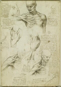
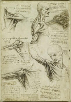
The superficial anatomy of the shoulder and neck; The muscles of the shoulder c.1510-11
Pen and ink with wash, over black chalk | 29.2 x 19.8 cm (sheet of paper) | RCIN 919003
-
A folio from Leonardo's 'Anatomical Manuscript A'.
Recto: Studies of the anatomy of the shoulder. Most of the drawings are based on study of a live subject, as informed by a knowledge of the underlying structures obtained by dissection. They display perplexing anatomical features seen on many other sheets of the period – a wide gap between the two heads of the sternocleidomastoid muscle on the sternum and clavicle, and a fascicular appearance to the muscles pectoralis major (from the chest to the arm) and deltoid (shoulder). For example, in the study at lower left we see a strongly contracted clavicular portion of pectoralis major and a virtually flaccid sternocostal portion, and the deltoid appears divided almost in half.
The drawing at the centre of the page shows the left shoulder from above with the subject facing to the left, whereas the drawing at centre left, which at first glance depicts the same region, shows the right shoulder from above with the subject facing to the right. The two images should thus mirror one another, but the musculature is very asymmetric: in the latter drawing, the deltoid is again divided and appears to receive a major contribution from the clavicular portion of pectoralis major.
The drawings to lower right show the structures underlying the superficial views. The omohyoid muscle is seen underneath and almost perpendicular to the sternocleidomastoid, though it seems to attach to the clavicle rather than to the scapula. The cephalic vein within the deltopectoral groove is well shown in all three drawings.
In a note Leonardo discusses his understanding of movement as composed of separable elements. He distinguished between ‘simple’ movements caused by one muscle or muscle group only; ‘mixed’ movements (elsewhere called compound or composite movements), caused by two independently operating muscles; and ‘decomposite’ or doubly compound movements, caused by three independently operating muscles. This is an expression of Leonardo’s understandable urge to resolve complex physical scenarios into simpler elements. His work as an anatomist led him to see that the apparently infinite movements of the body were the result of a finite number of muscle actions.
*
Verso: Studies of the muscles of the right shoulder. At upper centre and lower right Leonardo divided pectoralis major into four fascicles, and pectoralis minor is thus visible beneath; in the neck one can see sternocleidomastoid, omohyoid, levator scapulae and trapezius. Trapezius perplexed Leonardo: it is composed of superior fibres that descend, middle fibres that are horizontal and inferior fibres that ascend, and he therefore sometimes rendered it as several distinct muscles.
The drawing at top left is very similar to that on RCIN 919001r, though here the deltoid is in place; that muscle is also composed of three distinct sets of fibres, and Leonardo thus rendered it in a number of different ways – here it is virtually split in two, with distinct heads on the acromion and spine of the scapula.
At centre left is the deep structure of the shoulder and scapula from the front, with the ribs lifted away (roughly sketched in a lighter ink to the right). The contribution of the broad sheet of subscapularis to the ‘rotator cuff’ of the shoulder is elegantly demonstrated. The long head of biceps is present in its entirety, and its short head and coracobrachialis are indicated by two of the ‘tabs’ on the coracoid process (the other two are pectoralis minor). Leonardo has correctly shown the long head of triceps attaching to the border of the scapula. A note records the function of teres major and latissimus dorsi in rotating the humerus, but their attachments should be adjacent anterior to posterior, not proximal to distal as illustrated.
The drawing below places the ribs over the scapula and adds another layer of muscle. A nerve, either the median or ulnar, runs adjacent to coracobrachialis and can be followed up into the neck, and the coracoclavicular ligament is clearly depicted. Most of these structures are seen again in the drawing at lower right.
At top right is perhaps the most complex of Leonardo’s ‘thread diagrams’, summarising the entire three-dimensional structure of the shoulder in a single depiction. All the major muscles, both anterior and posterior, are reduced to threads or cords along their lines of force. The clavicle and ribs are also reduced in thickness to improve the ‘transparency’ of the diagram, but as Leonardo acknowledges in the note tucked under the arm, the density of information in such a diagram hinders its intelligibility, and he reminds himself to draw the diagram larger while maintaining the same thickness of ribs and muscle-threads.
Text from M. Clayton and R. Philo, Leonardo da Vinci: Anatomist, London 2012Provenance
Bequeathed to Francesco Melzi; from whose heirs purchased by Pompeo Leoni, c.1582-90; Thomas Howard, 14th Earl of Arundel, by 1630; Probably acquired by Charles II; Royal Collection by 1690
-
Creator(s)
Acquirer(s)
-
Medium and techniques
Pen and ink with wash, over black chalk
Measurements
29.2 x 19.8 cm (sheet of paper)
Other number(s)

