-
1 of 253523 objects
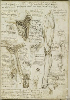
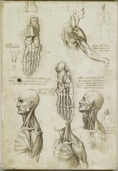
The throat, and the muscles of the leg (recto); The bones of the foot, and the muscles of the neck (verso) c.1510-11
Black chalk, pen and ink, wash | 29.0 x 19.6 cm (sheet of paper) | RCIN 919002

Leonardo da Vinci (1452-1519)
The throat, and the muscles of the leg (recto); The bones of the foot, and the muscles of the neck (verso) c.1510-11


-
A folio from Leonardo's 'Anatomical Manuscript A'.
Recto: a study of the lower left limb of a man, with the muscles of the thigh and calf emphasised; seven studies of the palate, tongue, larynx and hyoid bone; studies of the tongue, mouth, larynx, trachea and pharynx; studies of the tongue, throat, larynx, trachea, main bronchi and oesophagus in profile to the left; seven details of the larynx.
The leg was presumably the first study to be made, but the remainder of the page is devoted to the throat, with studies of the pharynx, larynx and trachea and their functions in breathing, speaking and swallowing. The large drawing at upper left displays many of the features seen in the subsequent, less extensive studies; the odd form of some of the structures may have been derived from animal dissection. Leonardo has shown the uvula, the wishbone-shaped hyoid bone, the thyroid gland, and the thyroid, cricoid and tracheal cartilages. But his understanding of the function of these structures was merely speculative: the thyroid gland, for instance, is ‘made to fill in where the muscles are missing, and … hold the trachea apart from the bone of the clavicle’.
The three drawings at centre and centre left show the palatoglossus and palatopharyngeus muscles pulling the hyoid bone backwards in order to press the epiglottis over the opening of the larynx during swallowing, thus preventing food or drink entering the larynx, as shown in the sketch at centre right. This is the reason that ‘one cannot swallow and breathe or speak at the same time.’
A similarly mechanical account of vocalisation is not easy (cf. RCIN 919115). Leonardo noted correctly that ‘the voice is generated at the head of the trachea’, and the frontal section of the larynx at lower centre shows the vocal cords and arytenoid cartilages forming a space (rima glottidis) referred to by Leonardo as a ‘flute’. He had studied vortices in wind and water, and he believed that turbulence in the expired air passing through the narrowed area of the rima glottidis set up vibrations that produced the voice (cf. his analysis of blood flow through the similarly shaped aortic valve on 919082-3 etc). This is substantially correct, with fine muscle movements adjusting the length and tension on the vocal cords to vary the air flow. But Leonardo inferred no role for the uvula in speech, regarding it as ‘the dripstone from which falls the humour that descends from above, and falls by way of the oesophagus to the stomach’. This ancient physiology, in which the systems of the body were controlled by the movement and balance of the ‘humours’, sits uncomfortably alongside the wealth of acute anatomical and physiological observations on the page.
*
Verso: the head and shoulders with emphasis on musculature; two studies of the bones of the foot; lateral movement of the big toe; three studies of the left side of a man; some studies of details and notes on the drawings.
The studies of the bones of the foot are mostly accurate, and the few inconsistencies may have resulted from Leonardo using fresh material, poorly cleaned dried material with fragments of ligament remaining, or dried material bound together too loosely. The drawing at the centre of the page shows the foot from below, and concentrates on the topography of the cuboid bone, with features labelled a, b and c – the attachment site of the short plantar ligament, the cuboid tuberosity, and the groove for the tendon of fibularis longus respectively.
The drawing at upper left shows a left foot, though the tibia and fibula hovering above are in the configuration for a right foot; Leonardo may have realised his error, as the lines drawn downwards from the fibula do not end on any tarsal bone. The little toe is shown with two phalanges – three is now usually stated as normal, but in fact two or three phalanges for the little toe occur in roughly equal numbers. The small diagram alongside indicates the action of the abductor and adductor muscles of the toes, those responsible for ‘lateral movement’, to use Leonardo’s term. Muscles analogous to those that move the fingers from side to side are found on the toes, but the ligaments and shape of the phalanges hinder the distinct actions, and usually the best we can do is spread the toes somewhat.
The four drawings along the bottom of the page concentrate on the muscles of the neck and shoulder, and bear some of the peculiarities found throughout Leonardo’s depictions of this region. The neck muscles, however, are beautifully presented. The two drawings to the left show the same stage of dissection, with all the muscles intact; in the drawing to the right sternocleidomastoid has been removed to display more clearly the complex of underlying muscles, nerves and vessels, most of which can be identified. The two bellies of the digastric muscle are shown again in the detail of the jaw from below, alongside.
Text from M. Clayton and R. Philo, Leonardo da Vinci: Anatomist, London 2012Provenance
Bequeathed to Francesco Melzi; from whose heirs purchased by Pompeo Leoni, c.1582-90; Thomas Howard, 14th Earl of Arundel, by 1630; probably acquired by Charles II; Royal Collection by 1690
-
Creator(s)
Acquirer(s)
-
Medium and techniques
Black chalk, pen and ink, wash
Measurements
29.0 x 19.6 cm (sheet of paper)
Other number(s)