-
1 of 253523 objects
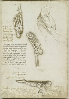
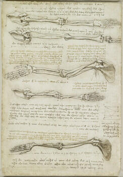
The bones of the foot; The bones and muscles of the arm c.1510-11
Black chalk, pen and ink, wash | 29.3 x 20.1 cm (sheet of paper) | RCIN 919000
-
A folio from Leonardo's 'Anatomical Manuscript A'.
Recto: a study of the trunk of a man, in profile to the right, with the right arm extended, indicating the surface muscles; the skeleton of a left foot; the same, seen from below; six diagrammatic drawings of the skeleton of the big toe; the bones of a left foot, in profile to the left and the corresponding leg bones, drawn upside down; notes on the drawings.
Verso: five studies of the skeleton of the right upper limb, showing pronation and supination; a diagram demonstrating pronation; notes on the drawings.
The upper two drawings show the arm and shoulder from above, held directly away from the side of the body with the palm facing upwards (supinated). In the first drawing the bones are in their natural positions, and in the second they are separated out to demonstrate their articulation. The biceps brachii muscle is beautifully illustrated, with its double origin (the name means ‘two heads of the arm’) on the scapula. Leonardo discovered that biceps has two actions, both bending the arm at the elbow and supinating the arm (turning the palm to face upwards). While there are other muscles whose sole purpose is supination, the biceps brachii is the strongest supinator of the forearm. It would be two centuries before Leonardo’s observation was repeated.
The third drawing gives a front view of the arm, with biceps cut away from its insertion on the radius and thus out of position, and two slips of pectoralis minor dangling from the coracoid process of the scapula. The small square marked between the ulna and radius near the wrist represents pronator quadratus, one of the two muscles primarily responsible for pronation (rotating the radius over the ulna to turn the palm downwards) – the position of the arm in the fourth and fifth drawings on the page. Leonardo concluded that as the bones cross and thus become oblique during pronation, the forearm must shorten a little (as illustrated in the small geometrical diagram in the right margin), though this is difficult to observe in practice. The other primary muscle of pronation is pronator teres, which is seen on the forearm in the final drawing. Like biceps, pronator teres has two heads, on the humerus and the ulna, though the latter attachment is not clearly shown.
Text adapted from M. Clayton and R. Philo, Leonardo da Vinci: Anatomist, London 2012Provenance
Bequeathed to Francesco Melzi; from whose heirs purchased by Pompeo Leoni, c.1582-90; Thomas Howard, 14th Earl of Arundel, by 1630; probably acquired by Charles II; Royal Collection by 1690
-
Creator(s)
Acquirer(s)
-
Medium and techniques
Black chalk, pen and ink, wash
Measurements
29.3 x 20.1 cm (sheet of paper)
Other number(s)

