-
1 of 253523 objects
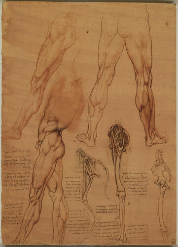
The leg muscles and bones of man and horse c.1506-8
Red chalk, pen and ink, on orange-red prepared paper | 28.2 x 20.4 cm (sheet of paper) | RCIN 912625
-
The muscles of a man’s legs, in several views, together with the bones of the pelvis and legs, with ‘cords’ indicating the lines of action of the muscles. At lower centre, a diagram of the same structures in the horse.
The superficial drawings of legs belong to a long series of similar studies executed by Leonardo around the time of his work on the Battle of Anghiari (1503-6 and later). They are life studies informed by his knowledge obtained by dissection, though how much human dissection he had carried out by this date is unclear. He notes ‘where the muscles are separated from one another you will draw in their borders’, and he has thus exaggerated their boundaries. But as Leonardo was not able to differentiate easily between the subcutaneous fat and the muscle itself, the shapes of some of the muscles appear rather odd, especially the gluteal muscles in the buttocks and gastrocnemius and soleus in the lower leg.
The two studies at lower centre compare the pelvic and leg bones of man and horse – Leonardo notes astutely that ‘to match the bone structure of a horse with that of a man you will have to draw the man on tip-toe in depicting his legs’. A few muscles are represented by threads: rectus femoris from the anterior iliac spine to the patella; tensor fasciae latae from roughly the same point towards the lesser trochanter of the femur (in the horse this trochanter is much more prominent than in the human); and the gluteal muscles, represented by a number of threads (two in the horse, four in the human) running from the iliac crest towards the greater trochanter. Leonardo had the same problem with the gluteal muscles as he did with the deltoid. The gluteals really are separate muscles, and usually the difference between gluteus maximus, g. medius and g. minimus is easily seen, but the more superior fibres of g. maximus can sometimes be confused with g. medius, and the cleavage fascia between g. medius and g. minimus continually confounded Leonardo.
The drawing at lower right has been called an ‘anatomical fantasy’, blending the bones of a horse with those of a man. It is much more likely that Leonardo intended it to be purely human, but incorporated errors (in particular the extended ischium below the coccyx) derived from his superior knowledge, at that date, of equine anatomy – the study of human bones and muscles to the left displays the same errors. Conversely Leonardo’s study of the horse here was to some degree compromised by his knowledge of human anatomy: the pelvis is too upright and not long enough, and the femur is too long and thin.
Text adapted from M. Clayton and R. Philo, Leonardo da Vinci: Anatomist, London 2012Provenance
Bequeathed to Francesco Melzi; from whose heirs purchased by Pompeo Leoni, c.1582-90; Thomas Howard, 14th Earl of Arundel, by 1630; probably acquired by Charles II; Royal Collection by 1690
-
Creator(s)
Acquirer(s)
-
Medium and techniques
Red chalk, pen and ink, on orange-red prepared paper
Measurements
28.2 x 20.4 cm (sheet of paper)
Markings
watermark: Flower (cut) [-]
Category
Object type(s)
Other number(s)


