-
1 of 253523 objects
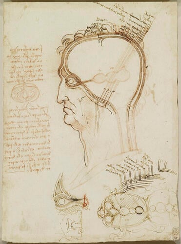
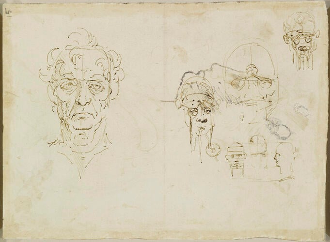
Recto: The layers of the scalp, and the cerebral ventricles. Verso: Studies of the head c.1490-92
Recto: Red chalk, pen and ink. Verso: Pen and ink, discoloured white, and faded metalpoint | 20.3 x 15.3 cm (sheet of paper) | RCIN 912603

Leonardo da Vinci (1452-1519)
Recto: The layers of the scalp, and the cerebral ventricles. Verso: Studies of the head c.1490-92

Leonardo da Vinci (1452-1519)
Recto: The layers of the scalp, and the cerebral ventricles. Verso: Studies of the head c.1490-92


-
Medieval physiology imagined the cerebral ventricles as three bulbous cavities that housed the mental faculties. The first was where the mind’s ‘raw material’ was gathered – the senso comune, imagination and fantasy; in the second this information was processed, through reasoning and so on; and the third was where the results were stored, in the memory. But Leonardo was unconvinced by this arrangement, repeatedly trying different layouts throughout his anatomical career, and finally abandoning the traditional system of the localisation of mental faculties. (See also RCIN 912626; 919057-9; 919127; 919070; 912602.)
The principal drawing here imagines the head sectioned through the middle, and in the left margin its layers are likened to those of an onion. Leonardo lists the layers in order: ‘hair; scalp; muscular flesh; pericranium arising from the dura mater; cranium, that is, bone; dura mater; pia mater; brain’. This is essentially repeated in the detail to lower right, with dura mater and pia mater transposed; while most of these layers correspond with modern usage, Leonardo used the term pia mater to refer to what is now called the arachnoid mater (named as such by Frederik Ruysch in 1664); what we now call the pia mater is practically inseparable from the underlying brain and neural tissue, and Leonardo would have been unable to differentiate it as a separate layer of the brain. He correctly shows the dura mater extending along the optic nerve to the posterior surface of the eye. Even the globe of the eye is comprised of layers, as indicated; the lens, however, is not located in the center of the vitreous body as shown, but immediately behind the iris.
While the shape and extent of the cranial cavity is not particularly accurate, Leonardo has prominently indicated the frontal sinus, one of his discoveries from sectioning the skull in 1489 (919057). The arrangement of the cerebral ventricles and sensory nerves is more conventional than in 912626, and corresponds closely to traditional images of the sensory apparatus.
The diagram at lower right shows the head sectioned horizontally at eye level and the crown of the head flipped back: all the sensory nerves, not just the optic, converge on the first ventricle. It must be supposed that this last diagram was imaginary rather than recording an actual sectioning of the head performed by Leonardo.
On the verso of the sheet is a drawing of an old man's head, full face, with various anatomical scribbles of a head and cranium, together with a rough sketch of a head in profile left, not by Leonardo.
Text adapted from M. Clayton and R. Philo, Leonardo da Vinci: Anatomist, 2012, no. 14.Provenance
Bequeathed to Francesco Melzi; from whose heirs purchased by Pompeo Leoni, c.1582-90; Thomas Howard, 14th Earl of Arundel, by 1630; probably acquired by Charles II; Royal Collection by 1690
-
Creator(s)
Acquirer(s)
-
Medium and techniques
Recto: Red chalk, pen and ink. Verso: Pen and ink, discoloured white, and faded metalpoint
Measurements
20.3 x 15.3 cm (sheet of paper)
Object type(s)
Other number(s)