-
1 of 253523 objects
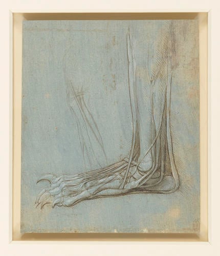
The anatomy of a bear's foot c.1488-90
Metalpoint, pen and ink, white heightening, on blue-grey prepared paper | 16.1 x 13.7 cm (sheet of paper) | RCIN 912372
-
During the latter part of the 1480s, Leonardo dissected the left hind leg of a bear. At that time bears were common in the mountains of Italy; they were hunted for sport and kept in captivity for entertainment, and Leonardo could thus easily have obtained a specimen for dissection. His particular interest in the animal was probably due to its plantigrade gait – like humans, bears walk with their feet flat on the ground (digitigrade animals such as dogs and cats walk on some portion of the digits, and unguligrade animals such as horses or cattle walk on the tip of a digit, usually in the form of a hoof). Leonardo’s dissection would therefore have given him an insight into the anatomy of the human foot at a time when he had little access to human material.
The drawing shows the bones, muscles and tendons of the lower leg and foot, in a lateral or outside view. Prominent are the extensor retinaculum ligament holding in place the tendons of extensor digitorum longus on the front surface of the ankle. (The omission of this ligament was to be an odd feature of Leonardo’s later studies of the human foot; on RCIN 919061r, a page of notes also of around 1510 he reminded himself to ‘make a discourse on the hands of each animal in order to show in what way they vary, as with the bear in which the ligament joins the tendons of the toes together over the neck of the foot’ - suggesting that he thought this ligament was peculiar to the bear.)
The tendons of fibularis longus, brevis and tertius pass behind the lateral malleolus (ankle bone), and the superficial fibular nerve can be seen running diagonally above the lateral malleolus and entering the dorsum or upper surface of the foot. The Achilles or calcaneal tendon appears to pass right around the calcaneus and along the sole of the foot (its insertion on the calcaneus is more correctly shown in 912375). The tendon of extensor digitorum longus, and muscles and tendons possibly of extensor digitorum brevis and/or an interosseous or lumbrical muscle, can also be seen inserting on the upper side of the toes.
Text adapted from M. Clayton and R. Philo, Leonardo da Vinci: Anatomist, London 2012, no. 7Provenance
Bequeathed to Francesco Melzi; from whose heirs purchased by Pompeo Leoni, c.1582-90; Thomas Howard, 14th Earl of Arundel, by 1630; probably acquired by Charles II; Royal Collection by 1690
-
Creator(s)
Acquirer(s)
-
Medium and techniques
Metalpoint, pen and ink, white heightening, on blue-grey prepared paper
Measurements
16.1 x 13.7 cm (sheet of paper)
Other number(s)

